- Grades 6-12
- School Leaders
At ISTE? Join us at booth 1359!
Every product is independently selected by (obsessive) editors. Things you buy through our links may earn us a commission.

30 Hands-On Circulatory System Activities for Kids
Make a working heart pump model, hold a stuffed animal “blood drive,” and more!

Kids need to learn how their bodies work and how to take care of them. Suffice it to say that the heart and circulatory system are a big part of that. Learning about the circulatory system through fun activities is hands-on—check your pulse to help students understand what’s happening—and it’s conceptual.
These hands-on, interactive activities teach and reinforce learning about the circulatory system. They’ll help kids learn how important it is to take care of their one and only heart!
1. Reveal the circulatory system

This is such a cool way to introduce the circulatory system to kids! Make DIY invisible ink using supplies from your kitchen and draw a model of the system. Then, let kids reveal it like magic, helping them envision what lies beneath their skin.
Learn more: Circulatory system reveal activity at Taming Little Monsters
2. Mold a Play-Doh circulatory system

Draw an outline of the human body, then grab some red and blue clay or play dough to make arteries, veins, and the heart itself.
Learn more: Play dough circulatory system at The Pinay Homeschooler
3. Watch a kid-friendly circulatory system video
YouTube has lots of videos that can help kids understand their hearts, blood vessels, and more.
We like this one from TED-Ed, which has free accompanying teacher materials .
4. Read a heart-healthy book

Check the library or buy one of these smart books for pre-K and elementary-age kids.
Buy it: The Heart by Seymour Simon; The Big Red Heart by Z.B. Tucker; Heart Your Heart by Paul Showers; A Drop of Blood by Paul Showers, all at Amazon
5. Play the Circulatory System Game

This free printable PDF game from Ellen McHenry is one of the most popular circulatory system activities around. Create a life-size body model, and spin your way around the system!
Learn more: Circulatory system game at The Fantastic Five
6. Turn a plastic bag into an inflatable heart

Make a simple heart model from a plastic bag, and use the straws to breathe into it and make it “beat” in rhythm.
Learn more: Circulatory system in a bag activity at Kids Activities Blog
7. Scoop water, and try to beat the clock

The heart pumps about 1.3 gallons of blood per minute. Think you can keep up? Fill a container with water, then set a timer. Use a small cup to scoop water into another container as fast as you can. Can you beat your own heart?
Learn more: Pumping blood activity at Primary Theme Park
8. Craft a simple stethoscope

Kids know that doctors use stethoscopes to listen to their hearts. Make a simple version from a cardboard tube and plastic funnel so kids can try it on their own.
Learn more: Stethoscope activity at Science Sparks
9. See your pulse using a marshmallow
Now that they’ve heard their heart, try this idea to see it in action. Push a toothpick into a marshmallow and set it on your upturned wrist. Hold very still and you should see the toothpick bounce up and down along with your pulse!
10. Build a functioning heart pump

Now it’s time to learn how the heart does its job. Use plastic bottles and drinking straws to make a working model that actually pumps “blood” from one chamber to the next.
Learn more: Heart activity at STEAM Powered Family
11. Tie yarn to learn about veins and arteries

The heart works with the veins, arteries, and capillaries to move blood around the body. Tie different colors of yarn together to represent the three and see how they all function together.
Learn more: Veins and arteries activity at The Fantastic Five
12. Engineer a complete circulatory system

Put it all together with this functioning circulatory system model. Get step-by-step instructions at the link.
Learn more: Circulatory system model at Do Science
13. Bottle of blood

Take a closer look at blood and learn about the different types of cells, platelets, and the plasma that they all float around in. Use your favorite candies to represent each in this easy model.
Learn more: Bottle of blood activity at My Joy-Filled Life
14. Explore different blood types with food coloring

Learning about blood types? This clever experiment with food coloring helps kids learn which types are compatible with each other. If the dyes stay the same color when mixed, the types are compatible. If they change color, then they’re not.
Learn more: Blood type experiment at Our Journey Westward
15. Test your blood type knowledge

In this interactive online game, kids “draw blood” from a patient, then run tests to find the blood type and perform a lifesaving transfusion. You can take a tutorial first to learn how it all works
Learn more: The Blood Typing Game at Educational Games
16. Hold a stuffed animal “blood drive”

This might be the cutest of all the circulatory system activities! Gather up some stuffies, assign them “blood types,” then hold a blood drive! Kids learn about blood types and the importance of being willing to donate blood.
Learn more: Toy blood drive at Highhill Education
17. Learn about the danger of narrowed veins and arteries

We talk a lot about “healthy foods,” but what makes some foods bad for your heart? Learn about cholesterol and its effects on veins and arteries with this effective demo.
Learn more: Arteries and veins activity via Teach Engineering
18. Exercise to keep your heart healthy

A healthy heart needs exercise too. Create a tic-tac-toe board with the free printables at the link, then toss a beanbag (bonus points for making heart-shaped ones!) to see which exercise you’ll do next.
Learn more: Heart-healthy exercises at Make and Takes
19. Host a Jump Rope for Heart event

The American Heart Association created Jump Rope for Heart and Hoops for Heart to raise awareness about the importance of heart health for kids. Students have a chance to earn money to support cardiovascular research too.
Learn more: Jump Rope for Heart activities at The P.E. Specialist
20. Label and fill in a heart anatomy coloring page

Teacherplanet.com has many coloring sheets that include opportunities for students to label each part of the heart and the respiratory system. Give your students some crayons or markers and let their creativity flow while simultaneously teaching them the parts of the heart.
Learn more: Heart coloring page at Teacher Planet
21. Watch a Bill Nye video about blood and circulation
What better way to engage your students than by turning on “Bill Nye the Science Guy”?! This video is very efficient in explaining the heart, delivered by a character students will recognize and love.
22. Make a heart and circulatory system poster

Separate your students into groups and give them 30 minutes to create as accurate a poster about the circulatory system as they can. Hang up the posters and keep them up while you finish your lesson plan, so your students can have a physical reference throughout the day.
Learn more: Circulatory system poster at Homeschool Den
23. Play the Circulation Game

This game teaches students about circulation, including the “map” of the circulatory system in and out of the lungs. Ellenjmchenry.com has detailed instructions on how to set up this engaging game involving small teams, perfect for the classroom.
Learn more: The Circulation Game at Ellen McHenry’s Basement Workshop
24. Graph your heart rate

Have students take their resting pulse and record it. Then, run a few laps, do 20 jumping jacks or dance for one minute, and record their heart rate again. Talk about why their heart rate changes and what happens.
Learn more: Graph heart rate activity at The Rocket Resource
25. Create a model of the heart

Another great use for play dough. Create a model of the heart and label the various parts.
Learn more: Heart model at Windhill 21
26. Blood sensory bin

Create a sensory bin with plasma, red blood cells, white blood cells, and platelets for students to discover.
Learn more: What is blood? sensory bin at I Can Teach My Child
27. Blood slime

Another way to show what goes into slime is with this blood slime. The slime is clear and filled with red blood cells, white blood cells, and platelets. For older kids, look at the ratio of each type of cell and incorporate that into the blood slime.
Learn more: Blood slime at Royal Baloo
28. Heart booklet

This cute booklet printable is a great way for students to take home what they’ve learned about the circulatory system.
Learn more: Heart booklet at Mrs. Jones’ Creation Station
29. Circulation system disc

This disc is a handy tool for studying the circulatory system.
Learn more: Circulatory system disc at Teaching in Room 6
30. Do the Circulation!
Do the Circulation with Schoolhouse Rock. Print out the lyrics and have students highlight opinions and facts. Then, talk about why authors might incorporate opinions into a song about circulation.

Learn more: Circulation analysis activity at Teaching in Room 6
Love these circulatory system activities? Check out 31 Interactive Ways To Teach Kids the Anatomy of the Human Body.
Plus, be sure to sign up for our newsletters to get all the latest teaching tips and more, straight to your inbox..

You Might Also Like

31 Interactive Ways To Teach Kids the Anatomy of the Human Body
Working hand models, edible spinal columns, and more! Continue Reading
Copyright © 2024. All rights reserved. 5335 Gate Parkway, Jacksonville, FL 32256
- Create new account
- Reset your password
Register and get FREE resources and activities
Ready to unlock all our resources?
Human circulatory system
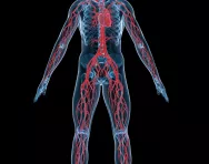
The circulatory system is one of the most important systems in the body. Made up of the heart, blood and blood vessels , the circulatory system is your body's delivery system. Your heart plays and important part in being healthy. It keeps all the blood in your circulatory system flowing. Blood helps oxygen get around your body. When you exercise you can feel your pulse, it tells you how fast your heart is pumping.
The body's circulatory system is responsible for transporting materials throughout the entire body. It delivers nutrients, water, and oxygen to your billions of body cells and carries away wastes such as carbon dioxide that body cells produce. It is an amazing highway that travels through your entire body connecting all your body cells.
At the centre of this system is the heart, an amazing organ. The heart beats about 3 billion times during an average lifetime . It is a muscle about the size of the fist. The heart is located in the centre of the chest slightly to the left. I ts job is to pump blood and keep the blood moving throughout the body . The blood is pumped around a complex network of blood vessels extending to every part of the body.
Blood carries the oxygen and nutrients needed to fuel the activities of the body’s tissues and organs , and it plays a vital role in removing the body’s waste products. An average-sized adult carries about 5 litres (9 pints) of blood.
Top 10 facts
- If you were to lay out all of the arteries, capillaries and veins in one adult, end-to-end, they would stretch about 60,000 miles (100,000 kilometres).
- It takes 20 seconds for blood to circulate the entire body . Oxygenated blood leaves the aorta at about 1 mile an hour.
- The power output of the heart ranges from 1-5 watts per minute, which is the equivalent to the usage of a 60 watt bulb. It has been said that enough energy is produced by the human body in a day to drive a truck 20 miles.
- Red blood cells live for up to 4 months and make approximately 250,000 round trips around the body before returning to the bone marrow, where they were born, to die.
- Between 2.5 and 3 million red blood cells (called erythrocytes ) are lost and replaced every second.
- Across the animal kingdom, heart rate is related to body size : in general, the bigger the animal, the slower its resting heart rate. An adult human has an average resting heart rate of about 75 beats per minute, the same rate as an adult sheep. But a blue whale's heart is about the size of a small car, and only beats five times per minute. A shrew, on the other hand, has a heart rate of about 1,000 beats per minute.
- The ancient Egyptians believed the heart, rather than the brain, was the source of emotions, wisdom and memory, among other things.
- After circulating within the body for about 120 days, a red blood cell will die from aging or damage. Bone marrow constantly manufactures new red blood cells to replace those that perish.
- The oxygen-rich blood that flows through your arteries and capillaries is bright red. After giving up its oxygen to your bodily tissues, your blood becomes dark red as it races back to your heart through your veins.
- “ Ventricle ” means “little belly".

Boost Your Child's Learning Today!
- Start your child on a tailored learning plan
- Complete the activities added each week
- Watch your child's maths & English confidence grow!
Did you know?
- We see and hear about hearts everywhere. A long time ago, people even thought that their emotions came from their hearts, maybe because the heart beats faster when a person is scared or excited. Now we know that emotions come from the brain, and in this case, the brain tells the heart to speed up.
- Your heart is a very strong muscle that pumps blood around your body. It is made of four chambers, two upper chambers and two lower chambers. Blood enters the upper chambers. These squeeze and push the blood into the lower chambers, which then squeeze and push the blood out of your heart.
- The heart works tirelessly – more than 2.5 billion times over an average lifetime – to pump blood around the body. The heart’s contractions or ‘squeezes’ are triggered by electrical impulses that come from a specialised area of heart tissue.
- Your pulse is a measure of how fast your heart is beating. It is the number of beats your heart makes in one minute. Your heart beats faster or slower depending on what you are doing. You can feel your pulse at certain points on your body. The easiest place to feel it is in your wrist, using the first two fingers of your other hand. When you sit, the average heart beats about 80 times per minute. However, everybody is different, so your pulse could be higher or lower than this.
- When you exercise , your heart beats more quickly. This is because your muscles are working harder and need more oxygen to keep going. Your lungs also work harder, making you breathe more quickly to get more oxygen. When you sleep, your muscles need less oxygen, so your heart slows down.
Circulatory system gallery:
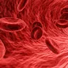
- Magnified red blood cells
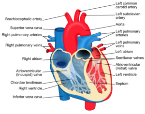
- A diagram of the human heart
- Blood vessels in the body
- A blood pressure check
- Doctors use stethoscopes to listen to heart and lung sounds
- A diagram of the circulatory system
The circulatory system is centred on the heart , an amazing organ that constantly works to pump blood around blood vessels in every part of the body. Blood carries all the things, like oxygen , that cells need to thrive and keep us healthy.
The heart is made up of four different blood-filled areas, and each of these areas is called a chamber . There are two chambers on each side of the heart. One chamber is on the top and one chamber is on the bottom.
The two chambers on top are called the atria . (If you're talking only about one, call it an atrium.) The atria are the chambers that fill with the blood returning to the heart from the body and lungs. The heart has a left atrium and a right atrium.
The two chambers on the bottom are called the ventricles . The heart has a left ventricle and a right ventricle. Their job is to squirt out the blood to the body and lungs. Running down the middle of the heart is a thick wall of muscle called the septum . The septum's job is to separate the left side and the right side of the heart.
The atria and ventricles work as a team — the atria fill with blood, then push it into the ventricles. The ventricles then squeeze, pumping blood out of the heart. While the ventricles are squeezing, the atria refill and get ready for the next contraction.
The blood moves through many tubes called arteries and veins, which together are called blood vessels . These blood vessels are attached to the heart. The blood vessels that carry blood away from the heart are called arteries . The ones that carry blood back to the heart are called veins .
The human body needs a steady supply of blood to keep it working right. Blood delivers oxygen to all the body's cells. To stay alive, a person needs healthy, living cells. Without oxygen, these cells would die. If that oxygen-rich blood doesn't circulate as it should, a person could die.
Remember that your heart is a muscle. In order for it to be strong, you need to exercise it. How do you do it? By being active in a way that gets you slightly out of breath, like skipping, dancing, or playing tennis or football. Try to be active every day for at least 30 minutes.
Words to know for the circulatory system:
Aorta - the main artery in mammals that carries blood from the left ventricle of the heart to all the branch arteries in the body except those in the lungs. Arteries - a blood vessel that is part of the system carrying blood under pressure from the heart to the rest of the body. Atrium - one of the upper chambers of the heart that takes blood from the veins and pumps it into a ventricle. Capillaries - an extremely narrow thin-walled blood vessel that connects small arteries arterioles with small veins to form a network throughout the body. Carbon dioxide - a heavy, colourless, odourless gas. Cells - the cell is the basic unit of life. Some organisms are made up of a single cell, like bacteria, while others are made up of trillions of cells. Human beings are made up of cells, too. Circulatory - relating to the circulation of the blood. Complex - made up of many interrelated parts. Contractions - a tightening or narrowing of a muscle, organ, or other body part. Nutrients - a substance that provides nourishment. Organ - a complete and independent part of a plant or animal that has a specific function. Oxygen - a colourless, odourless gas that is essential for plant and animal respiration. Perish - to come to an end or cease to exist. Pulse - the regular expansion and contraction of an artery, caused by the heart pumping blood through the body. Transporting - to carry somebody or something from one place to another. Veins - a blood vessel that carries blood to the heart. Waste - unwanted or unusable remains, or by-products.
Related Videos
Just for fun...
- Try some circulatory games and puzzles
- Complete a heart and circulatory system quiz
- Download free circulatory system quizzes, puzzles and memory games from Curiscope
- A heart to print out and label
- Try the heartbeat calculator
- Play the Blood Typing game and find out more about blood types
Best kids' books about the circulatory system and the heart

Find out more
- A kids' guide to the heart and blood
- Watch BBC Teach video about the circulatory system
- Download a factsheet and worksheet about blood and the circulatory system and find out how and why people donate blood
- Heart and Circulatory system quiz
- The role of blood in the circulatory system
- See inside the human body and find out more about the circulatory system
- Watch a medical video of how the chambers of the heart contract and relax to push blood through the circulatory system
- Watch a BBC Bitesize video about how our circulatory system keeps us alive
See for yourself
- Watch a cartoon that describes how the heart works
- See exactly how your heart pumps blood throughout your body
Give your child a headstart
- FREE articles & expert information
- FREE resources & activities
- FREE homework help
In order to continue enjoying our site, we ask that you confirm your identity as a human. Thank you very much for your cooperation.
- Success stories
- Spine and back
- Pelvis and perineum
- Head and neck
- Neuroanatomy
- Cross sections
- Radiological anatomy
- Types of tissues
- Body systems

Register now and grab your free ultimate anatomy study guide!

Circulatory (cardiovascular) system
Author: Niamh Gorman, MSc • Reviewer: Francesca Salvador, MSc Last reviewed: September 12, 2023 Reading time: 34 minutes
/images/vimeo_thumbnails/258779944/Ynw0qC4YypuucQulRKAbNg_overlay.jpg)
The circulatory system, also called cardiovascular system , is a vital organ system that delivers essential substances to all cells for basic functions to occur. Also commonly known as the cardiovascular system, is a network composed of the heart as a centralised pump, blood vessels that distribute blood throughout the body, and the blood itself, for transportation of different substances.
The circulatory system is divided into two separate loops: The shorter pulmonary circuit that exchanges blood between the heart and the lungs for oxygenation; and the longer systemic circuit that distributes blood throughout all other systems and tissues of the body. Both of these circuits begin and end in the heart.
| Functions | Transport of gases, nutrients, electrolytes, wastes, hormones |
| Heart | - myocardium, endocardium, epicardium - left and right atria, left and right ventricles - arteries (oxygenated blood), veins (deoxygenated blood) |
| Blood vessels | Arteries, veins, capillaries Hierarchy: Heart -> arteries -> arterioles -> capillaries [gas exchange - oxygenated blood becomes deoxygenated] -> venules -> veins -> heart |
| Circulations | - superior and inferior vena cava (with deoxygenated blood) -> right atrium -> right ventricle -> right and left pulmonary artery -> capillaries of each lung (oxygenation of the blood) -> pulmonary veins -> left atrium -> systemic circulation - left atrium -> left ventricle -> aorta and all of its branches -> capillaries -> veins -> superior and inferior vena cava -> pulmonary circulation - ascending aorta -> right coronary artery -> right marginal branch, posterior interventricular artery, left coronary artery -> anterior interventricular branch (anastomoses with the posterior branch), circumflex artery |
| Blood | with (red blood cells) - contain hemoglobine and carry oxygen throughout the blood vessels (white blood cells) - immune system cells (platelets) - coagulation cells |
| Clinical relations | Arteriosclerosis, cerebrovascular disease, peripheral artery disease, aneurysm, varices, arrhytmia, heart failure |
This article will explain everything that is important about the circulatory system, as well as the clinical relations to it.
Pulmonary circulation
Systemic circulation, coronary circulation, portal system, shunts and anastamoses, erythrocytes (red blood cells), leukocytes (white blood cells), thrombocytes (platelets), vascular diseases, cardiac diseases, blood disorders.
The main function of the circulatory (or cardiovascular) system is to deliver oxygen to the body tissues, whilst simultaneously removing carbon dioxide produced by metabolism. Oxygen is bound to molecules called haemoglobin that are on the surface of the red blood cells in the blood.
Beginning in the heart , deoxygenated blood (containing carbon dioxide) is returned from systemic circulation to the right side of the heart . It is pumped into pulmonary circulation and is delivered to the lungs , where gas exchange occurs. The carbon dioxide is removed from the blood and replaced with oxygen. The blood is now oxygenated, and returns to the left side of the heart .
Have you already learned the basic anatomy of the heart? Test your knowledge with our heart diagrams, quizzes and worksheets.
From there, it is pumped into the systemic circuit, delivers oxygen to the tissues , and returns again to the right side of the heart . The blood also acts as an excellent transport medium for nutrients, such as electrolytes, as well as hormones. The blood also transports waste products, that are filtered from the blood in the liver.
The heart is a muscular pump that is the central component of the circulatory system. It is divided into a right and left side by a muscular septum . The muscular component of the heart, the myocardium , is composed of involuntary cardiac muscle . It is lined by a membrane called the endocardium internally, as well as an external epicardium . Contraction of the cardiac muscle cells is stimulated by electrical impulses that are sporadically fired from the regulatory centres of the heart: the sinuatrial node in the roof of the right atrium , and the atrioventricular node in the septum between the atria and the ventricles . The sinuatrial node is widely regarded as the natural pacemaker of the heart.
The heart is continuously going through a series of contractions and relaxations. Systole refers to when the ventricles of the heart simultaneously contract, diastole is when the ventricles relax. During systole, blood is forcibly pumped out of the ventricles into the outflow tracts of their corresponding circulation. The atria are filling with blood at the same time. During diastole, the ventricles are relaxed, and blood flows from the atria into the corresponding ventricles.
Deoxygenated blood from systemic circulation returns to the right atrium via the superior and inferior vena cava . The coronary sinus , returning blood from the coronary circulation, also opens into the right atrium. The blood in the right atrium flows into the right ventricle through the right atrioventricular valve ( tricuspid valve ) during diastole. During systole, the right ventricle contracts, directing the blood into the conus arteriosus at the base of the pulmonary trunk . Contraction of the ventricle causes the tricuspid valve to shut, preventing backflow of blood into the right atrium. Between the conus arteriosus and the pulmonary trunk is a valve; the pulmonary valve . In diastole, the valve closes to prevent backflow of blood into the right ventricle.
The pulmonary trunk splits into a right and a left pulmonary artery , serving the right and left lung respectively. Deoxygenated blood flows into the capillaries of each lung, where it is then oxygenated. The pulmonary veins collect the newly oxygenated blood from the lung, and return it to the left atrium, where it will be passed into systemic circulation.
Oxygenated blood enters the left atrium from the pulmonary circulation via the pulmonary veins . During diastole, blood passes from the left atrium to the left ventricle through the left atrioventricular valve ( bicuspid valve ). In systole, the left ventricle contracts, forcing blood into the aorta . The blood passes through the false into the ascending aorta .
The ascending aorta becomes the arch of the aorta , where three large arteries branch from it: the brachiocephalic trunk , the left common carotid artery and the left subclavian artery. These arteries supply oxygenated blood to the head and neck , and to the upper limbs .
The descending aorta is the continuation of the arch of the aorta inferiorly. In the thorax it is referred to as the descending or thoracic aorta , and gives off numerous branches in the thorax.
The latter passes into the abdominal cavity through the diaphragm through the aortic hiatus at the level of T12. From there, it is referred to as the abdominal aorta . The abdominal aorta gives branches to the structures in and surrounding the abdominal cavity, and terminates by bifurcating into the common iliac arteries , which will supply the pelvic cavity and lower limbs .
The branches of the aorta passes towards their intended structures, with branching occurring along their length. The terminal branches enter the tissues, and pass towards the capillary beds of the tissues in vessels called arterioles . Gas exchange occurs between the blood and the tissues. The blood is collected from the capillaries by venules , which unite to form the veins of the systemic circulation. These veins ultimately drain to the right atrium via the superior and inferior venae cavae.
The coronary circulation refers to the blood supply to the heart itself. It is a component of the systemic circulation . The right and left coronary arteries branch directly from the ascending aorta, immediately above the aortic valve. The right coronary artery passes to the right and gives off two main branches: the right marginal branch along the right border of the heart and the posterior interventricular ( posterior descending ) artery, which descends along the interventricular septum on the base of the heart.
Learn everything about the coronary arteries and veins with the following study unit and quiz.
:format(jpeg)/images/study_unit/coronary-arteries-and-cardiac-veins/WPXMtQtGZlik50VQJfSEQ_Coronary_vessels.png "homework for circulatory system")
The left coronary artery passes to the left, and gives off the anterior interventricular ( Ieft anterior descending ) artery which descends on the anterior aspect of the interventricular septum to anastamose with the posterior interventricular artery at the apex of the heart. It also gives off the circumflex artery .
The venous drainage of the heart is achieved by the coronary sinus , which drains the main veins of the heart:
- the great cardiac vein ,
- the middle cardiac vein , and
- the small cardiac vein , which drains directly into the right atrium.
The portal system is the system of veins that drain the blood from the intestines and directs it to the liver to be filtered. The superior and inferior mesenteric veins , draining the jejunum down as far as the upper rectum , along with the splenic vein draining the spleen, pancreas , and stomach , unite to form the hepatic portal vein , which empties blood into the liver. Toxins are filtered out by the liver, and the filtered blood is returned to the inferior vena cava via the hepatic veins.
Types of blood vessels
Arteries carry blood away from the heart. They have thick walls and a narrow lumen , to resist the high pressure from the blood being forced out of the heart. As the arteries travel toward the more peripheral tissues, they begin a process of segmentation, decreasing in diameter and wall thickness with each division. The major arterial outflow tracts of the heart are the aorta (systemic), and the pulmonary trunk (pulmonary). The coronary arteries are the arteries that supply oxygenated blood to the tissues of the heart itself.
Arteries are typically divided into three types:
- distributing arteries that transport blood to specific organ systems, with a high muscular component in their walls;
- the small and muscular resistance vessels or arterioles
Pressure in these arteries decrease from its highest level in the conducting arteries to the lowest in the arterioles. The walls of the arteries are divided into 3 layers: the tunica intima (internal), the tunica media (middle) and the tunica externa (external).
For descriptive purposes, it is easiest to describe the types of blood vessels in the sequence that they occur as they pass from the heart to the peripheral tissues, and form the peripheral tissue back to the heart.
How's your knowledge of the major arteries of the cardiovascular system? Our cardiovascular system diagrams, quizzes and free worksheets are the best way to find out.
Types of arteries
Large elastic arteries : are the conducting arteries and examples include the aorta and its main branches; the brachiocephalic trunk, the left common carotid artery, the left subclavian artery and the terminal common iliac arteries. These carry blood from the heart to the smaller conducting arteries. The pressure in the these arteries is at the highest level of the entire circulatory system. The tunica intima is lined by endothelium and the tunica media has a large elastic component . Muscular arteries : are the distributing arteries and contain a large proportion of smooth muscle in their tunica media. They are lined internally by endothelium. The tunica externa is composed of fibromuscular connective tissue , with a larger proportion of elastic fibres than collagen contributing to the elasticity of this layer in the muscular arteries.
Arterioles : are the connecting vessels between the muscular arteries and capillary beds of the organs. They have small endothelial cells with nuclei projecting into the lumen of the vessel, a thin muscular wall about two layers thick, and a thin tunica externa. They control the flow of blood into the capillaries by contraction of the smooth muscle in the tunica media, which acts as a sphincter. Capillaries : are the closest vessels to the organs. Their walls measure one large endothelial cell in thickness and provide the only barrier between the blood and the interstitial fluid of the tissues. They have a narrow lumen which is just thick enough to allow the passage of the largest blood cells. The permeability of capillaries varies depending on the surrounding tissues and the type of junctions between the adjacent endothelial cells in the vessels wall.
Types of veins
Venules : are formed when two or more capillaries converge. They are lined by flat endothelial cells and a thin tunica externa. These are called postcapillary venules. The muscular component appears in venules as their lumen increases, producing muscular venules. Veins : are formed with the union of muscular venules. In comparison to arteries, veins have a relatively thin wall and a larger lumen . The structure of the walls is similar to that of arteries, but a considerably smaller amount of muscle is present in the tunica media of veins. Veins are capacitance vessels , meaning they have a distensible wall and can expand to accommodate large volumes of blood.
Most peripheral veins have structures called valves , which are projections of the tunica interna into the lumen of the vessel. Valves prevent the backflow of blood through the veins, by passively closing when the direction of flow of the blood reverses. Valves are absent in the veins of the thorax and abdomen . The overall hierarchy of blood vessels follows this order: arteries → arterioles → capillaries → venules → veins.
So now you know the types of blood vessels - but what about their histological features? Learn and test your knowledge at the same time using our blood vessels diagrams and artery and vein quizzes.
Arteries form connections between each other called anastomoses, which creates a continuous supply of blood throughout different areas. In the event of occlusion of an artery to a specific area, blood supply can be maintained to the tissue via the anastomosis with an artery of an adjacent area.
A direct anastomosis occurs where two arteries are joined directly to each other, such as in the radial and ulnar arteries via the palmar arches. Convergence anastomoses occur where two arteries unite to form a single artery, as in when the vertebral arteries join to form the basilar artery . A transverse anastomosis is where a small artery connects two larger arteries, for example, the anterior communicating artery connecting the right and left anterior cerebral arteries .
Connections between the arterial and venous systems are present throughout the body. For example, in the mesentery , metarterioles can connect the arterioles to venules, and blood can either flow into or bypass the capillary beds. Control of this flow is by local demand of the individual tissues.
Arteriovenous anastomoses are a direct connection between small arteries and small veins. These occur in regions such as the skin of the nose, lips and ears , in the mucosa of the alimentary canal, and nasal and oral cavities .
A portocaval anastomosis occurs where there is a connection between the systemic and portal system of veins. These occur at venous plexuses, such as around the oesophagus , the umbilicus , and the rectum.
The blood is the mobile component of the circulatory system. Blood is bright red when oxygenated and dark red/purple when deoxygenated. Blood consists of a cellular component suspended in a liquid called plasma.
Plasma is a clear fluid that accounts for approximately 55% of blood, and is composed of over 90% water. Plasma contains a high concentration of electrolytes , such as sodium, potassium and calcium. Also dissolved in plasma are plasma proteins . These include clotting factors, mainly prothrombin, immunoglobulin, polypeptides and other protein molecules, and hormones.
Erythrocytes are the most abundant of blood cells, accounting for approximately 99% of all blood cells. They are biconcave disc shaped cells that lack a nucleus. Erythrocytes have a globulin protein called haemoglobin on their surface for oxygen to bind to. The proportion of red blood cells to plasma is called the haematocrit . Measured as a percentage, it is used as a reference point for the oxygen carrying capacity of a person; when there is a higher percentage of red blood cells present, more haemoglobin is present to carry oxygen.
Aged erythrocytes are ingested by macrophages in the liver and spleen . The iron released in the breakdown of the erythrocytes is used to synthesise new erythrocytes, or is stored in the liver as ferritin .
Blood Grouping
Antigens are present on the surface of erythrocytes, and can react with antibodies causing agglutination of the red blood cells. This is the basis of the ABO blood grouping system . Individuals inherit two alleles, one from each parent, that code for a specific blood group. Blood groups can be homozygous , where the alleles are the same, or heterozygous where alleles are different:
| Allele | |
| AA | A |
| BB | B |
| OO | O |
| AB | AB |
| AO | A |
| BO | B |
Specific blood groups have antibodies that are sensitive to the alleles absent from their erythrocytes. For example, blood group A will carry the A antigen and the anti-B antibodies.
These are divided in 5 groups: monocytes, lymphocytes , neutrophils , basophils and eosinophils . These groups are distinguishable from each other by cell size, shape of nucleus and cytoplasm composition. These groups can themselves be grouped into 2 groups: granulocytes and agranulocytes . This classification is based on the presence or lack of granules in the cytoplasm of the cell. Collectively, white blood cells form part of the immune response .
Granulocytes
Neutrophils, eosinophils and basophils fall into this category of white blood cells. Leukocytes are classified into this group based on the presence of vesicles, called granules, in their cytoplasm. Granulocytes are largely involved in inflammatory and allergic responses .
Neutrophils : are the most abundant white blood cells, accounting for about 40-75% of all leukocytes. The number of neutrophils varies, and increases in response to acute bacterial infections. They have an irregular, segmented nucleus. They mainly function in the defence of the body against microorganisms, and can ingest foreign substances by phagocytosis . They are also involved in inflammation. Neutrophils have a short life span, spending 4-7 hours in circulation and a few days in connective tissue.
Eosinophils : are similar to neutrophils, but are far fewer in number. Their nucleus is prominently bilobed, and the granules in the cytoplasm are large. Their motility mirrors that of other leukocytes, and they migrate from the circulation into the tissues. They increase in number in allergic reactions, and play a prominent role in the defense against parasites . They are only weakly phagocytotic, involved more so in the breakdown of particles too large for phagocytosis. The circulate for approximately 10 hours, and spend a few days in the tissues.
Basophils : are the smallest of the granulocytes. They are small in number, accounting for 0.5-1% of all leukocytes. They are distinguishable by the large, clearly visible granules in their cytoplasm. Their nucleus is irregular shaped, and sometimes bilobed, but is often obscured by the granules. The granules are membrane bound vesicles containing a variety of inflammatory agents. These vesicles herniate, dumping their contents and triggering immediate allergic hypersensitivity , such as seen in reactions like hay fever. The dumping of these agents also triggers the migration of other granulocytes to the area.
Agranulocytes
Monocytes and lymphocytes fall into this category due to the absence of granules in their cytoplasm. They are also referred to as mononuclear leukocytes, referring to the presence of a single lobed nucleus.
Monocytes : are the largest leukocytes in relation to physical size. They account for 2-8% of all leukocytes. They typically have large uni-lobed nuclei with a characteristic indentation on one side. Monocytes are phagocytic cells . Circulating monocytes transition into macrophages when they migrate from the circulation to the tissues.
Lymphocytes : are the second most abundant leukocyte, accounting for 20-30%. They are the only white blood cell that can re-enter circulation having migrated to the tissues. They are variable in size and lifespan: some live merely days, others are long-lived, and are involved in immunological memory . Lymphocytes are divided into two types: B-lymphocytes and T-lymphocytes.
B-lymphocytes synthesize and secrete antibodies specific to foreign molecules. They also stimulate other non-lymphocytic leukocytes to phagocytose. B-lymphocytes are involved in adaptive immunity , and produce memory B cells, which remain in the body and are activated in response to a specific antigen.
T-lymphocytes develop and mature in the thymus , then migrate to and are stored in secondary lymphoid organs. They are involved in the ongoing immunity of the cell, with their function not solely dependent on the response to an antigen. T-lymphocytes are divided into three subgroups. Cytotoxic T cells directly target infected cells; Helper T cells direct destruction by recruitment of other immune cells; and Regulatory T cells are involved in developing the tolerance of cells to an antigen.
Platelets are small, irregular shaped cells that lack a nucleus. They are present in large numbers and have highly adhesive properties. Platelets are highly involved in haemostasis . They are activated in the event of damage to a blood vessel. They accumulate at the site of injury and essentially plug the wound. Following adherence at the site of injury, platelets and the surrounding tissues release factors that trigger a complex sequence of events. A clot is formed to close the wound. The clot is then retracted and the edges of the wound are pulled together to close it and repair the vessel. Platelets circulate in the blood for approximately 10 days, before they are removed from the blood by macrophages .
Want some practice identifying blood cells? Then try the quiz below!
Clinical notes
Diseases affecting the cardiovascular system are collectively referred to as cardiovascular diseases. Vascular diseases relate to the blood vessels. Cardiac diseases affect the heart itself. Hematologic diseases are those of the blood. Diseases of the cardiovascular system can be congenital (present since birth) or acquired (related to age, diet, lifestyle and predisposition).
Arteriosclerosis is the thickening of the walls of arteries, reducing function. Atherosclerosis is a specific form of arteriosclerosis, where plaque builds up on the endothelium of arteries, causing them to narrow and reducing oxygen delivery to the tissues.
Coronary artery disease occurs in the arteries supplying the heart itself, with narrowing of the coronary arteries causing reduced oxygen delivery to the heart tissue. This can result in a condition called angina , which is essentially spasming of the coronary arteries due to reduced blood flow. Myocardial Infarction (heart attack) is also caused by the narrowing of the coronary arteries due to atherosclerosis. A myocardial infarction occurs when the artery becomes completely occluded due to dislodged plaque or development of a thrombus (blood clot).
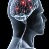
Cerebrovascular disease affects the arteries supplying the brain . One of the most common presentations is ischemic stroke , which is also caused by atherosclerosis. Ischemic stroke results in a reduced blood flow to brain regions, leading to impaired brain function. It can be caused by the development of a thrombus or the passing of an embolus (blockage causing substance) from another region of the body to the cerebral circulation.
Peripheral artery disease is reduced blood flow to the limbs due to atherosclerosis.
An aneurysm is a localised weakening in the wall of a blood vessel. It can result in bulging of the vessel wall. Thrombus formation and embolisation can also occur. Aneurysms can rupture, leading to significant blood loss depending on where they occur. Particularly lethal sites of aneurysm formation are in the abdominal aorta, the circle of Willis in cerebral circulation, and in the renal vessels.
Varices occur where blood vessels become enlarged and twisted. They can occur at multiple sites in the body. One of the most prominent sites of varices is in the veins of legs, termed varicose veins. Other common sites of varices are at sites of portocaval anastamoses, such as esophageal varices, umbilical varices (caput medusae) and anorectal varices (hemorrhoids or piles).
Cardiovascular diseases can also solely affect the heart. Cardiomyopathy is a collection of diseases that affects the heart muscle. The muscle can become enlarged (hypertrophic) and rigid, causing decreased heart function, arrhythmias (irregular heart rate), and sometimes even heart failure .
The valves of the heart can also be affected by disease. There are two main types: valve incompetence , in which the valve is unable to function sufficiently; and valve stenosis , where the orifice between the valve narrows as the valve is unable to open fully. Mitral valve disease affects the mitral valve that lies between the left atrium and ventricle. It is normally caused by a combination of valve incompetence and stenosis. Aortic valve disease affects the aortic valve, and is largely caused by stenosis of the valve with contribution from regurgitation, which is backflow through the valve.
Inflammation of the heart tissues can also occur. It includes inflammation of the inner endocardium ( endocarditis ) and the middle muscular layer ( myocarditis ). Pericarditis is the inflammation of the pericardium , which comprises the outer layer of the heart itself and the pericardial sac which encloses the heart in the thoracic cavity.
Congenital heart diseases
Congenital heart diseases are those which have been present since birth. They are largely present as left to right shunts , where blood is shunted from areas of higher pressure to areas of lower pressure. Oxygenated blood is passed back to the right side of the heart and mixed with deoxygenated blood. Such shunts can go unnoticed in a number of patients, while others may require surgical intervention.
An atrial septal defect occurs when blood is shunted from the left atrium (higher pressure) to the right atrium (lower pressure) through an opening in the interatrial septum . This opening usually results from the failure of an embryological shunt, the foramen ovale, to close after birth. This defect is specifically referred to as a patent foramen ovale . A ventriculoseptal defect is when an opening in the interventricular septum allows blood to pass from the left ventricle into the right ventricle.
Another embryological shunt exists near the heart in the embryo, shunting blood from the pulmonary trunk into the aorta. This is called the ductus arteriosus , and pressure changes after birth usually force this opening to shut. A patent ductus arteriosus occurs when the ductus does not close after birth, and allows blood to flow from the higher pressure arch of the aorta into the lower pressure pulmonary trunk.
These are disorders affecting the components of the blood. They can largely be divided depending on which of the blood cells they affect.
Anemia is a blood disorder affecting red blood cells . Patients suffering with anemia have a decreased oxygen carrying capacity due to a decrease in the number of red blood cells, or a reduced amount of haemoglobin in the blood. There are multiple different types of anemia, some of which are the following:
- Iron deficient anemia is the most common form of anemia. It is the result of insufficient intake of iron, an increase in the amount of iron lost, or inadequate absorption of iron. Women are more likely to be affected by this from of anemia due to menstruation and the higher demands of iron placed on their body during pregnancy.
- Megaloblastic anemia is caused by a decrease in the intake or absorption of vitamin B12 or folic acid. This results in the production of large, insufficient red blood cells.
- Perniciou s anemia is the result of insufficient hemopoiesis, or production of red blood cells by bone marrow.
- Hemorrhagi c anemia is caused by loss of red blood cells through excessive bleeding.
- Aplasti c anemia occurs due to the destruction of red bone marrow , which leads to a reduction in the number of red blood cells being produced.
- Sickl e cell anemia is a condition in which the shape of the red blood cells is altered into a sickle shape. These cells cannot easily pass through capillaries and tend to clump together, blocking the blood vessel. They are also prone to rupturing, with their rapid break down resulting in a reduced oxygen carrying capacity.
Leukemia refers to a group of cancers affecting the red bone marrow . Theses cancers cause abnormal white blood cells to multiply uncontrollably, which interferes with normal red blood cell, white blood cell and platelet production. This results in a decrease in oxygen carrying capacity, susceptibility to infection, and abnormal clotting. Leukemia spreads easily from the bone marrow to the lymph nodes , liver and spleen, causing them to enlarge. Symptoms are caused mainly by disruption to the production of other blood cells, including fatigue, pale skin and cold intolerance that is usually observed in anemia.
There are two methods of classification of leukemia. The first is based on the presentation of the disease: Acute leukemia refers to those that have developed rapidly; Chronic leukemia develops over an extended period of time. The second classification is based on the type of cells affected: Lymphoblastic affects lymphoid stem cells; Myelogenous affects myeloid stem cells. Thus, there are four types of leukemia:
- Acute lymphoblastic leukemia is the most common form of the disease occurring in children, though it can also affect adults as well.
- Acute myelogenous leukemia is found in both adults and children.
- Chroni c lymphoblastic leukemia is usually present in adults, especially those over the age of 55.
- Chronic myelogenous leukemia usually affects adults.
Treatment of leukemia involves methods such as chemotherapy, radiation therapy, stem cell transplantation and blood transfusion among others.
Thrombocytopenia
This is a disorder of the thrombocytes , or platelets. It results in a low number of platelets in the blood. Patients with this disorder are prone to excessive bleeding and may experience frequent nose bleeds or bleeding gums, as well as excessive bruising.
This is an inherited blood disorder that causes spontaneous bleeding or bleeding where only minor trauma has occurred. It is caused by deficiencies of different clotting factors and can vary significantly in severity.
Reference List:
- F. Netter: Atlas of Human Anatomy, 6th Edition, Elsevier Saunders (2014).
- G.J. Tortora, B. Derrickson: Principles of Anatomy & Physiology, 13th Edition, Wiley (2012).
- J.A. Gosling, P.F. Harris, J.R. Humpherson et al.: Human Anatomy, Colour Atlas and Textbook, 5th Edition, Mosby Elsevier (2008).
- M.H Ross, W. Pawlina: Histology: A Text and Atlas, Wolters Kluwer, 7th Edition (2016).
- R. Drake, A.W. Vogl, A.W.M. Mitchell: Gray’s Anatomy for Students, 3rd Edition, Churchill Livingston Elsevier (2015).
Illustrators:
- Overview of the heart in situ (ventral view) - Yousun Koh
- Aortic arch (ventral view) - Yousun Koh
- Overview of coronary arteries and cardiac veins - Yousun Koh
- Anastamosis between superior mesenteric artery and inferior pancreatic artery (ventral view) - Esther Gollan
- Coronary circulation in a cadaver - Prof. Carlos Suárez-Quian
Circulatory (cardiovascular) system: want to learn more about it?
Our engaging videos, interactive quizzes, in-depth articles and HD atlas are here to get you top results faster.
What do you prefer to learn with?
“I would honestly say that Kenhub cut my study time in half.” – Read more.

Learning anatomy isn't impossible. We're here to help.

Learning anatomy is a massive undertaking, and we're here to help you pass with flying colours.
Want access to this video?
- Curated learning paths created by our anatomy experts
- 1000s of high quality anatomy illustrations and articles
- Free 60 minute trial of Kenhub Premium!
...it takes less than 60 seconds!
Want access to this quiz?
Want access to this gallery.
Circulatory and Cardiovascular System STEM
The circulatory system is one of the most important systems in the human body. It’s responsible for transporting blood, nutrients, and oxygen to all of our cells. Keeping it healthy is essential for overall health and wellness.
This STEM lesson plan on the circulatory system is the perfect way for students to learn about this vital system and how to keep it healthy. Through a variety of engaging activities and hands-on experiments, they’ll gain a thorough understanding of its structure and function. They’ll also discover how important it is to other body systems and learn practical tips for keeping it healthy.
Description
Additional information, what our circulatory and cardiovascular system stem lesson plan includes.
Lesson Objectives and Overview: Circulatory and Cardiovascular System STEM teaches students about the structure and functions of this body system. Students will be able to explain how and why to keep the circulatory system healthy. They will also discover how it works with other body systems to function properly. This lesson is for students in 4th grade, 5th grade, and 6th grade.
Classroom Procedure
Every lesson plan provides you with a classroom procedure page that outlines a step-by-step guide to follow. You do not have to follow the guide exactly. The guide helps you organize the lesson and details when to hand out worksheets. It also lists information in the yellow box that you might find useful. You will find the lesson objectives, state standards, and number of class sessions the lesson should take to complete in this area. In addition, it describes the supplies you will need as well as what and how you need to prepare beforehand. For this lesson, you will need red and green markers, water, large containers, and measuring cups. You might also choose to get a timer for the activity.
Options for Lesson
In the “Options for Lesson” section of the classroom procedure page, you will see some suggestions for additional activities or ideas to add to the lesson if you want to. One idea is to have students calculate the mean, median, mode, and range of the heart rates of the students in the class. (You may benefit from pairing this lesson with a math lesson related to central tendency and graphing.) Students could change the amount of water or size of the measuring cup to reflect different stages in life—child versus adult—during the practice. Consider talking about different animals and their hearts and how they differ from those of humans. Another option is to have a veterinarian come to the class to talk about the different circulatory systems of animals and compare them to humans.
Teacher Notes
The teacher notes page provides an extra paragraph of information to help guide the lesson. You can use the blank lines to write down any other ideas or thoughts you have about the topic as you prepare.
CIRCULATORY AND CARDIOVASCULAR SYSTEM STEM LESSON PLAN CONTENT PAGES
Introduction to the heart.
The Circulatory and Cardiovascular System STEM lesson plan has three pages of content. Students will first learn what this body system actually is. The circulatory system comprises the heart, blood vessels, and blood. There are two circulatory systems in the human body: pulmonary circulation and systemic circulation. With pulmonary circulation blood loops from the heart to the lungs and back. And with systemic circulation, it loops from the heart to all the other parts of the body and back.
With each heartbeat, blood travels throughout the body to carry oxygen and nutrients to the cells. Without blood, we wouldn’t be alive! This is why the heart has to pump blood continuously through the body. The heart is the main organ in the circulatory system. It is a muscle about the size of a fist on the left side of the chest. Its job is to propel blood throughout the body.
Each minute, the heart beats anywhere from 60 to 100 times, but it can go even faster than that if needed. The body sends the heart signals to pump more (or less) blood depending on the situation. For example, exercising or being scared tells the heart to pump faster to get more oxygen. But when we sleep, our body tells the heart to slow down. Each day, the heart beats around 100,000 times. That’s 30 million times a year and over 2.5 billion times over an average lifespan.
Parts of the Heart
The heart has four chambers: two ventricles and two atria. The bottom two chambers are the ventricles, which are separated by a wall called the interventricular septum. Ventricles are responsible for pumping blood out of the heart. The upper two chambers of the heart are the atria. Atria have the opposite job as ventricles and receive blood as it enters the heart. The wall between them is the interatrial septum. In addition, four sets of valves in the heart ensure that the blood can only flow in one direction.
Blood enters the heart through the right atrium, passes down to the right ventricle through the tricuspid valve, and flows through the pulmonary artery. It flows through the pulmonary artery to the lungs, where it gathers oxygen and removes carbon dioxide. The aorta is the main artery that takes away blood. Something called the mitral valve separates the left atrium and left ventricle, taking away blood from the heart.
Blood Vessels
Next, students will learn about blood vessels. Blood vessels constantly circulate blood throughout the body. As the blood circulates, it picks up nutrients from food and drops them off to the cells that need energy. Then it takes the waste from the cells (carbon dioxide) and drops it off in the lungs so we can breathe it out.
Blood vessels are tubes that run through the entire body to help carry the blood. Some of these are veins, which bring blood to the heart. Others are arteries, which take blood away from the heart. Arteries have to be super strong and thick because there is more pressure on them from the heart’s pumping. Veins don’t have to be as wide because they carry the blood back to the heart.
Our blood consists of red blood cells, among other things. Red blood cells carry oxygen, which is why it looks red. Blood also contains platelets that help our blood clot when we get a cut. In addition, it has white blood cells, which are responsible for killing germs and keeping the blood free from invaders. The red blood cells, white blood cells, and platelets float around in a substance called plasma. Together they make up our blood.
There are four main types of blood—A, B, O, and AB. Each type is slightly different from the others because of antibodies and antigens. If a person needs a blood donation, they must receive blood that matches their blood type. If the blood types do not match, the person receiving the blood can get very sick.
Keeping the Circulatory System Healthy
The circulatory system works with all the other systems in the body to help supply nutrients and remove waste. Our heart is an essential part of the system as it pumps blood throughout our bodies. It is necessary to keep it healthy and working well for our whole life.
Like all muscles, we can strengthen the heart through regular exercise. For example, a brisk 10-minute walk can increase the blood flow through the heart and make it stronger. Avoiding foods that are heavy in fat and sugar can also help our circulatory system remain healthy. On the flip side, eating healthy foods and snacks like natural fruits and vegetables can strengthen our heart and circulatory systems.
Excessive stress can hurt the circulatory system because it increases the levels of chemicals in the bloodstream that injure the cells. Deep-breathing exercises and learning to manage stress are part of a heart-healthy plan. Infections can be hard on the circulatory system as well. A good habit is to wash the hands often to reduce the chances of getting sick from the flu or other diseases.
CIRCULATORY AND CARDIOVASCULAR SYSTEM STEM LESSON PLAN WORKSHEETS
The Circulatory and Cardiovascular System STEM lesson plan includes three worksheets: an activity worksheet, a practice worksheet, and a homework assignment. Each one will help students solidify their grasp of the material they learned throughout the lesson. You can refer to the classroom procedure guidelines to know when to hand out each worksheet.
PULSE RATE ACTIVITY WORKSHEET
For the activity, students will collect data on their own pulse rate. First, they will put their hands on either their neck or wrist and count their pulse for 10 seconds. After multiplying that number by 6 to get their beats per minute, they will record the answer in the table. Students will then run in place as fast as they can for 15 seconds. Again, they will count their pulse for 10 seconds, multiply by 6, and record the answer in the table.
The worksheet lists four prompts regarding the data students collected about their pulse. They must explain why the rate changed after exercising and describes things that make a pulse rate change. Then they will list three activities to increase their pulse rate and two that decrease it.
On the second worksheet page, students will collect the heart rate data from four other students and fill in their information in the table. Finally, they will create a bar graph showing each students resting pulse rate in green and active pulse rate in red.
CIRCULATORY AND CARDIOVASCULAR SYSTEM STEM PRACTICE WORKSHEET
The practice worksheet requires students to see if they can work faster than their hearts. Students will first read a short passage at the top of the page about how much blood the heart pumps through their body every day. They will fill a container with one gallon of water and set a timer for one minute. Then they will use a small cup to scoop water into another container as fast as possible. Another students will count the scoops. At the bottom of the worksheet are four prompts for students to respond to.
EXERCISES HOMEWORK ASSIGNMENT
For the homework assignment, students will mark off spots on a tic-tac-toe board as they complete the exercise on that spot. They should do as many of each exercise as they can for two minutes. Then they will check their heart rate after each exercise and record the beats per minute in the box. Exercises include jumping jacks, push-ups, squats, and jogging in place.
Worksheet Answer Keys
The lesson plan document provides answer keys for the three worksheets. All the correct answers are in red to make it easier for you to compare them with students’ work. Some of the prompts on various worksheets are objective, so students’ answers will vary. The answer keys often provide sample responses in these cases. If you choose to administer the lesson pages to your students via PDF, you will need to save a new file that omits these pages. Otherwise, you can simply print out the applicable pages and keep these as reference for yourself when grading assignments.
| grade-level | 4th Grade, 5th Grade, 6th Grade |
|---|---|
| subject | Science |
| State Educational Standards | NGSS.MS-LS1-1, NGSS.MS-LS1-2, NGSS.MS-LS1-3 Lessons are aligned to meet the education objectives and goals of most states. For more information on your state objectives, contact your local Board of Education or Department of Education in your state. |
Thank you for submitting a review!
Your input is very much appreciated. Share it with your friends so they can enjoy it too!
A great resource!
My class loves the Learn Bright videos. They are professionally done and cover all the relative information on the topics.
Related products

Sports: Ultimate

Clouds STEM

Sports: Field Hockey
Make your life easier with our lesson plans, stay up-to-date with new lessons.
- Lesson Plans
- For Teachers
© 2024 Learn Bright. All rights reserved. Terms and Conditions. Privacy Policy.
- Sign Up for Free
Login Get started
- Extra Credit
- Uncategorized
The Circulatory System
- Teaching Staff
- March 18, 2019
- No Comments
There are multiple organ systems in the human body that work together to keep us alive. One of these systems is the circulatory system. The major components of the circulatory system are blood, blood vessels, and the heart. The main function of the circulatory system is to transport nutrients and gases throughout the body so cells can be provided with the nutrients and gases they need to function. It does this using blood and blood vessels , which act as a transportation network. Blood is made up of several components:
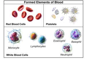
Red Blood Cells (44%) – contains hemoglobin, a protein that is iron rich, and allows for the red blood cells to carry oxygen
White Blood Cells (<1%) – part of the body’s immune system. They help to defend against infection and disease
Platelets (<1%) – responsible for blood clotting (thickening). This helps to prevent blood loss at the site of wounds.
Blood is contained inside the body in blood vessels. If you can imagine the body as a house, and blood as water, then blood vessels are the pipes carrying water throughout the home.
Types of Blood Vessels
Arteries – Carry blood that’s being pumped AWAY from the heart. This blood is under high pressure, and so arteries have thick, muscular walls.
Veins – Carry blood TO the heart. They contain valves which prevent the blood from flowing backwards. Veins have thinner walls than arteries as they are not under as much pressure.
Capillaries – Connect arteries and veins. They are very thin, and have walls made of epithelial tissue one cell layer thick. Because their walls are so thin, nutrients and gases can diffuse (travel) in and out of them. Capillaries can be found with alveoli (in the lungs; part of the respiratory system), and with the villi (in the stomach, part of the digestive system)
The circulatory system transports nutrients and gases throughout the body. Without it, the body’s cells would be without what they need to survive. One of the diseases that affects the circulatory system is high blood pressure or hypertension. Blood pressure is the pressure or force of blood as it flows through the circulatory system. Blood pressure is related to the volume of blood, heart rate, artery size and elasticity, and blood viscosity.
High Blood Pressure
Having high blood pressure puts strain on the heart because it’s forced to beat more rapidly. This can cause the heart to get bigger and become less efficient over time. One factor that can increase the risk of high blood pressure is a poor diet. High fat diets can cause the buildup of cholesterol in the arteries, which can cause them to become narrowed and blocked. As there is now less space for the blood to flow through (ie. artery is less wide), blood pressure increases.
In conclusion, the circulatory system’s main function is to transport nutrients and gases around the body via blood and blood vessels. The heart provides the pumping action to move the blood around. One disease of the circulatory system is high blood pressure, where the pressure of blood in blood vessels is higher than normal.
Sch oolTutoring Academy is the premier educational services company for K-12 and college students. We offer tutoring programs for students in K-12, AP classes, and college. To learn more about how we help parents and students in Aloha, Oregon visit: Tutoring in Aloha, Oregon.

Forces in Physics
The pythagorean theorem.
If you're seeing this message, it means we're having trouble loading external resources on our website.
If you're behind a web filter, please make sure that the domains *.kastatic.org and *.kasandbox.org are unblocked.
To log in and use all the features of Khan Academy, please enable JavaScript in your browser.
Biology archive
Course: biology archive > unit 33.
- The lungs and pulmonary system
- Red blood cells
Circulatory system and the heart
- Components of blood
Want to join the conversation?
- Upvote Button navigates to signup page
- Downvote Button navigates to signup page
- Flag Button navigates to signup page

Video transcript
- Science & Math
- Sociology & Philosophy
- Law & Politics
- The Human Circulatory System
1. transport oxygen and carbon dioxide 2. distribute nutrients and transport of wastes 3. maintenance of body temperatures 4. circulation of hormones
Made up of three components:
1. A fluid in which materials are transported (blood) 2. vessels in which the fluid moves (blood vessels) 3. a pump (the heart)
Circulatory system is actually two systems:
Pulmonary circuit :
-the right side of the heart pumps blood to the lungs – blood picks up oxygen and releases carbon dioxide – blood returns to the heart, on the left side
Systemic circuit :
– the left side of the heart pumps blood to all parts of the body – blood delivers oxygen and other nutrients – blood returns to the heart, on the right side
– two pumps, each pump is separated by the septum ; a muscle wall – the right pump, pumps to the pulmonary circuit – the left pump, pumps to the systemic circuit -each pump is made up of an atrium and a ventricle – atria receive blood from veins and pump into ventricles – ventricles receive blood from atria and pump blood into arteries – heart valves prevent blood from flowing backwards in the heart
Heart Rhythms, Sounds and Pressure
– the sinoatrial node sets the heart rhythm, about 72 beats per minute – the lubb-dubb heart sounds are caused by the closing of the valves – diastole is when the heart is relaxed, fills up with blood (semilunar valves close) – systole is when atria push blood into the ventricles, then the ventricles push blood into the arteries (AV valves close)
Related Posts
- Ontario Human Rights Code
- Heart Rate and Terms
- Human Genome Project
- Circulatory System: Function, Parts, Role
- Systems of the Human Body
Leave a Reply Cancel reply
Your email address will not be published. Required fields are marked *
Save my name, email, and website in this browser for the next time I comment.
Post comment
By signing up, you agree to our privacy policy .
Sign Up for our FREE Newsletter!
- Lesson Plans
- Lesson Templates
- Certificates
- Find Grants
- Fundraising
Search for Resources
You are here
Circulatory system lesson.
The lesson will begin with the teacher engaging the students with a presentation of "How the Blood Gets Around the Body" following a think quest presentation that covers the parts and functions of the circulatory system, including the brain, veins and arteries, heart and blood. Students will explore blood vessels by watching a short video clip, conducting a hands-on investigation about blood pressure. Next the teacher will lead a discussion and explain about the human heart and will use a "Map of the Human Heart" to show the class exactly how the heart pumps blood throughout your body and learn facts about the human heart. Students will get a chance to elaborate by creating a color picture of blood flow to, through and from the heart in their notebooks. To evaluate the students, they will watch a short video clip about the circulatory system and take the accompanying quiz.
Copyright © 2001 - 2024 TeacherPlanet.com ®. All rights reserved. Privacy Statement and Disclaimer Notice

Sign up for our free weekly newsletter and receive
top education news, lesson ideas, teaching tips, and more!
No thanks, I don't need to stay current on what works in education!
- STEM Ambassadors
- School trusts
- ITE and governors
- Invest in schools
- STEM careers inspiration
- Benefits and impact
- Our supporters
- Become a STEM Ambassador
- Request a STEM Ambassador
- Employer information
- Training and support
- STEM Ambassadors Partners
- Working with community groups
- Search icon
- Join the STEM Community
Human Circulatory system
Students need to be able to describe the human circulatory system, including the relationship with the gaseous exchange system. They will need to explain how the structure of the heart and blood vessels are adapted to their functions. Students need to know the components of blood and be able to explain how red blood cells, white cells, platelets and plasma are adapted to their functions.
Students often find it difficult to explain what is meant by double circulation, when delivering this topic ensure that students are able to clearly explain how blood is pumped to the lungs and body on each circuit and that it goes through the heart twice in each circuit. Asking students to give a two minute presentation on this (select different students at the start of different lessons), will help them articulate this
It is important when discussing/demonstrating the structure of the heart (for example in dissections) that students are shown the coronary arteries that supply the cardiac muscle, so that they appreciate that it is the blockage of these arteries that can cause heart attacks. There os often a misconception that it is the vessel which supply the heart which are blocked
The valves in the heart chambers are an important feature of the circulatory system so it is worth spending some time ensuring students appreciate the role that the valves play in the system, they should be a feature of any dissection.
Whilst this list provides a source of information and ideas for experimental work, it is important to note that recommendations can date very quickly. Do NOT follow suggestions which conflict with current advice from CLEAPSS, SSERC or recent safety guides. eLibrary users are responsible for ensuring that any activity, including practical work, which they carry out is consistent with current regulations related to Health and Safety and that they carry an appropriate risk assessment. Further information is provided in our Health and Safety guidance.
ABPI interactive website *suitable for home teaching*
This interactive website has a number of sections which are relevant to this topic area of GCSE Biology, these include: •
- The need for a transport system
- The circulatory system
- The blood vessels
- The blood and blood clotting
- Blood pressure
- Cardiovascular disease
- Prevention and treatment of cardiovascular disease.
In each section there is information and a brief self-test quiz. These sections could be used for independent learning/homework activities. Different students could be asked to complete specific tasks to answer in class having worked through the website for homework , tasks could include:
- What materials does blood transport throughout the body.
- Why do these materials need to be carried?
- Describe the role of each of the components in the blood.
- Compare and contrast the structures and functions of arteries, veins and capillaries.
- Explain why humans have a double circulatory system.
- Describe the structure of the heart.
- Describe the sequence of events in a cardiac cycle (heartbeat).
- How does the pressure change in each of the chambers during one heartbeat?
- Describe blood clotting and how this helps to prevent disease.
- Suggest ways that a person could help reduce their risk of having a heart attack
Nuffield Advanced Biology Topic Review: Circulation
Quality Assured Category: Biology Publisher: Nuffield Foundation
Although this is quite a dated publication, that was originally intended for 16-19 year old students, there are a range of sections within this publication which could be used as reading material with students.
Students could be provided with an on-line (or paper version) of a section and either individually or working in pairs, highlight key words and phrases, annotate the text to pick out important points and then provide a short half page summary of what they have read or they could read a section and then prepare a Powerpoint presentation to summarise what they have read.

Let's Dissect - the Heart
Quality Assured Category: Science Publisher: University of Bristol
This video is a detailed and graphic account of the dissection of a pig’s heart. The dissection, and accompanying commentary, shows all of the main features of the heart. It should be shown after students have been made aware of the heart structure.
The video could be used as the prelude for an actual dissection of a heart. There are also slides which highlight the position, structure and function of the main parts of the heart, including chambers, vessels, valves and heart wall. A diagram of the circulatory system allows students to watch a blood cell as it travels around the double circuit.
This resource has been designed as an independent study package, but is probably most useful as a teacher led activity There are obviously a number of issues and sensitivities related to dissection which teachers should always be aware of.
Heart Surgery and Dissection
Quality Assured Category: Science Publisher: Biotechnology and Biological Sciences Research Council (BBSRC) - UKRI
Produced by the Biotechnology and Biological Sciences Research Council (BBSRC), these materials help students to understand more about how the heart works and also includes a step-by-step guide to dissecting a heart obtained from a butcher.
In the first part of the resource, various aspects of cardiac biology are described and these include the use of magnetic resonance spectroscopy imaging techniques, studying skeletal muscle cells in vitro and the implanting of biomedical devices. The resource then describes in detail the dissection of a heart, obtained from a butcher, which allows the students to:
- identify the internal and external anatomy of a heart
- dissect a heart and be able to model the techniques of a heart surgeon
- discuss heart diseases and disorders, describe how they occur, and name risk factors and possible preventative measures
Electrocardiagrams
Quality Assured Category: Science Publisher: Institute of Physics
Although Electrocardiograms are not a requirement on GCSE Biology specifications, this resource provides a useful context into which required knowledge and understanding of the cardiovascular system can be framed.
The resource includes an animated PowerPoint which shows how blood flow and muscle contraction within the heart is related to the ECG trace. The graphs show how heart rate can be calculated by measuring the period between successive peaks on the ECG. There is also a worksheet for students to complete. This resource would be best used as a teacher led whole class activity, working through the PowerPoint and worksheet.

Off the Blocks
Quality Assured Category: Science Publisher: University of York Science Education Group (UYSEG)
This is a relatively dated publication, but it contains a range of potentially useful resources for teachers. The publication investigates various aspects of physiology in a sport related context, which will appeal to many students.
The publication is organised into "lessons" with teachers notes and activites/worksheets for students towards the end of the publication. Lessons 5,8 and 9 would all be useful for this GCSE Biology topic. The worksheets on pages 86,87 and 98 (as well as many others) could be used both within class or as homeworks without any amendments.
The Bloodmobile
An amusing video which may help some students remember the functions of blood cells.
- Fundamentals NEW
- Biographies
- Compare Countries
- World Atlas
cardiovascular system
Related resources for this article.
- Primary Sources & E-Books
Introduction
The human cardiovascular system is composed of the heart and the blood vessels—arteries, veins, and capillaries. Its purpose is to provide nutrients and oxygen to the tissues and to remove wastes from them. It is also where the body fights infections. The cardiovascular system is part of the circulatory system, which also includes the lymphatic system .
The human heart is a pear-shaped muscular organ about the size of a fist. A wall of muscle, the septum, divides the right side from the left. Each of these two sides is further divided into an atrium, or upper chamber, and a ventricle, or lower chamber. The human heart beats 60 to 80 times a minute while a person is at rest. The heart rests only about 0.4 second between beats.
Circulation of blood consists of two main circuits. Pulmonary circulation carries blood from the heart to the lungs where waste gases, mostly carbon dioxide, are removed from the blood, and oxygen is taken on by hemoglobin in the erythrocytes, or red blood cells. The blood then returns to the heart and is pumped to other parts of the body in the systemic circulation, which comprises the blood supply to the entire body except the lungs. Within the systemic circulation is the portal circulation, which supplies blood to the liver . Capillaries and veins carrying nutrient-rich blood from the digestive organs merge to form the portal vein, leading to the liver. Blood from the liver reenters the systemic circulation via the inferior vena cava.
Bright red, oxygen-rich blood from the pulmonary circulation flows from the left atrium into the left ventricle. With each beat of the heart, blood is forced through the large artery of the heart, called the aorta, and throughout the body by a network of arteries. The large arteries of this network are the carotid arteries in the neck and head, the axillary arteries in the arms, the abdominal arteries in the abdomen, and the external iliac arteries in the legs.
Numerous smaller arteries branch off these major ones. Each branching yields smaller and smaller arteries until the blood passes from the smallest arteries into the arterioles. These tiny structures connect to venules, forming a structure called a capillary. It is here that most of the blood-tissue exchange occurs, with the blood giving up its oxygen, nutrients, and fluid to the tissues, and the tissues giving up carbon dioxide and fluid wastes to the blood.
Several organs interact with the cardiovascular system in very specific ways. Nutrients and fluids move from the digestive organs into the blood. The spleen then filters the blood, removing old blood cells and microorganisms. Phagocytes, a type of white blood cell, engulf these materials and destroy them. The liver also filters the blood and detoxifies wastes such as ammonia into less toxic urea. The liver adds fibrinogen and prothrombin to the blood; these two proteins are essential for clotting. The kidneys extract water, minerals, urea, and other waste products from the blood. These materials are later excreted in the urine.
The blood that travels into the veins from the capillaries is oxygen-poor and dark red. The veins, like the arteries, branch into larger and larger structures and eventually return to the heart via the inferior vena cava and the superior vena cava. The blood enters the heart at the right atrium. The heart pumps it to the right ventricle; from there it is again pumped to the lungs, and the process of circulation repeats.
The arteries are the thickest of all blood vessels and have muscular walls that contract to keep the blood moving away from the heart. The veins are not muscular, but they contain valves to prevent the blood from flowing backward.
Circulation in the Fetus
The cardiovascular system of the human fetus differs from the adult system in several respects. Because the fetal lungs and digestive system are not yet functioning, these areas are bypassed by the fetal circulation. Instead, the mother’s oxygenated blood is carried through the uterus to the placenta.
The placenta unites the fetus to the uterus of the mother and serves the functions of nutrition, respiration, and excretion. The umbilical vein, which is wrapped around the umbilical cord, delivers the blood to the fetus. After it circulates through the fetus, the blood returns to the placenta via the umbilical arteries. At birth the fetal system shuts down and the circulation is rerouted to include the lungs and digestive system in the newborn infant.
Blood Pressure and Pulse
Arteries near the surface of the skin can be used to measure blood pressure, which reflects how the cardiovascular system is functioning. Blood pressure is governed by five elements—the strength of the heartbeat, the volume of blood, the viscosity of the blood, the resistance of the arterioles, and the elasticity of the arterial walls. An abnormality of any of these can cause the blood pressure to be too high or too low.
Blood pressure is measured by wrapping the cuff of an instrument called a sphygmomanometer around the arm just above the elbow. With a stethoscope over the brachial artery at the inside of the elbow, the rushing sounds of blood through the artery can be heard. The blood pressure, written as a fraction and measured in millimeters of mercury, is usually 100 to 120 over 60 to 80 in a resting, healthy person. The higher number represents the minimum pressure needed to stop completely the flow of blood. This is called the systolic blood pressure. The lower number represents the maximum pressure at which the beats heard through the stethoscope change from loud to soft—that is, when full blood flow is restored. This is called the diastolic blood pressure.
Another measurement of the circulation is made by taking the pulse. With the fingers (not the thumb) over the radial artery inside the wrist, the number of heartbeats is counted for six seconds and then multiplied by 10 to determine the heart rate, which is reported in beats per minute. In healthy adults the heart beats around 80 times per minute However, exercise and stress will cause both the pulse and the blood pressure to rise temporarily. Other pulse points of the body are the temple, side of the neck, back of the knee, top of the foot over the ankle, and groin.
Disorders of Circulation
Because the cardiovascular system is necessary for life, diseases that affect any part of it can have serious or fatal consequences. Hypertension, or high blood pressure, can be caused by an increase in the volume of blood pumped by the heart or by increased resistance to blood flow in the arterioles. Several diseases, including kidney disease, may cause hypertension; in some individuals, however, the cause may be unknown. Called the “silent killer,” hypertension is often symptomless but can lead to heart disease and stroke.
Arteriosclerosis is a group of diseases of the arteries. The diseases differ in some respects, but all are characterized by abnormal thickening and hardening of the walls of the arteries. The arteries become less elastic and cannot carry blood effectively throughout the body. The most common types of arteriosclerosis is atherosclerosis, a condition in which fatty deposits of cholesterol form in the arteries. These deposits can break away and travel to the heart or brain, causing heart attacks or strokes. Diets that are high in certain fats called saturated fats encourage the production of these deposits. ( See also anatomy, human ; disease, human .)
William A. Check
It’s here: the NEW Britannica Kids website!
We’ve been busy, working hard to bring you new features and an updated design. We hope you and your family enjoy the NEW Britannica Kids. Take a minute to check out all the enhancements!
- The same safe and trusted content for explorers of all ages.
- Accessible across all of today's devices: phones, tablets, and desktops.
- Improved homework resources designed to support a variety of curriculum subjects and standards.
- A new, third level of content, designed specially to meet the advanced needs of the sophisticated scholar.
- And so much more!
Want to see it in action?
Start a free trial
To share with more than one person, separate addresses with a comma
Choose a language from the menu above to view a computer-translated version of this page. Please note: Text within images is not translated, some features may not work properly after translation, and the translation may not accurately convey the intended meaning. Britannica does not review the converted text.
After translating an article, all tools except font up/font down will be disabled. To re-enable the tools or to convert back to English, click "view original" on the Google Translate toolbar.
- Privacy Notice
- Terms of Use
IMAGES
VIDEO
COMMENTS
delivers blood from the heart to the body tissues and back to the heart. systolic pressure. arterial pressure during the contracting phase of the cardiac cycle. thrombin. main coagulation enzyme. Thrombocytes. medical term for platelets. Study with Quizlet and memorize flashcards containing terms like ABO, Antecubital, aorta and more.
The main function of the circulatory system is to provide oxygen, nutrients and hormones to muscles, tissues and organs throughout your body. Another part of the circulatory system is to remove waste from cells and organs so your body can dispose of it. Your heart pumps blood to the body through a network of arteries and veins (blood vessels).
Three principal categories of blood vessels. • Arteries. • Carry blood away from heart. • Veins. • Carry blood back to heart. • Capillaries. • Connect smallest arteries to smallest veins to create a circuit. The Vessel Wall. Walls of arteries and veins have three layers called tunics.
The circulatory system transports oxygen and _____ to the body's cells. nutrients. The outer wall of the heart is surrounded by a sac called the: c. pericardium. When a stethoscope is used to listen to the heartbeat, two distinct sounds can be heard. This is called: b. lub, dub.
16. Hold a stuffed animal "blood drive". Highhillhomeschool.blogspot.com. This might be the cutest of all the circulatory system activities! Gather up some stuffies, assign them "blood types," then hold a blood drive! Kids learn about blood types and the importance of being willing to donate blood. Learn more: Toy blood drive at ...
The circulatory system is one of the most important systems in the body. Made up of the heart, blood and blood vessels, the circulatory system is your body's delivery system. Your heart plays and important part in being healthy. It keeps all the blood in your circulatory system flowing. Blood helps oxygen get around your body.
The cardiovascular system is a vital organ system which is quite literally at the centre of everything. Comprised of the heart, blood vessels and the blood itself, it is divided into two loops which both begin in the heart. The pulmonary circuit is responsible for exchanging blood between the heart and lungs for oxygenation, while the systemic circuit directs blood to the other tissues of the ...
Blood is made from four parts: Red blood cells which transport oxygen. White blood cells which protect against disease. Blood platelets which help the blood to clot and form scabs to repair a cut ...
Get comprehensive homework help for Circulatory System! Browse through questions students have asked on Circulatory System and see how Flexi helped them with answers and clear explanation. ... Pulmonary circulation is the part of the circulatory system that carries blood between the heart and lungs. Oxygenated blood leaves the right ventricle ...
Get help with your Circulatory system homework. Access the answers to hundreds of Circulatory system questions that are explained in a way that's easy for you to understand. ... In the circulatory system. bicarbonate leaves the red blood cells and enters the plasma through an exchange mechanism with _____ to maintain electroneutrality. (a ...
Circulatory system. The circulatory system, also called cardiovascular system, is a vital organ system that delivers essential substances to all cells for basic functions to occur. Also commonly known as the cardiovascular system, is a network composed of the heart as a centralised pump, blood vessels that distribute blood throughout the body, and the blood itself, for transportation of ...
The Circulatory System The circulatory system is an essential part of our body. 'Circulatory' means something that is going on a continuous circuit. This is exactly what is happening in our bodies all the time. Did You Know...? • In the average person, the heart beats about 2,500,000,000 times during a lifetime.
The circulatory system works with all the other systems in the body to help supply nutrients and remove waste. Our heart is an essential part of the system as it pumps blood throughout our bodies. ... Cardiovascular System STEM lesson plan includes three worksheets: an activity worksheet, a practice worksheet, and a homework assignment. Each ...
Study with Quizlet and memorize flashcards containing terms like What does the circulatory system transport?, what are the two main divisons of the circulatory system, the fluid connective tissue is _____,the_____ are blood vessels,and the_____is the heart and more.
The major components of the circulatory system are blood, blood vessels, and the heart. The main function of the circulatory system is to transport nutrients and gases throughout the body so cells can be provided with the nutrients and gases they need to function. It does this using blood and blood vessels, which act as a transportation network.
All animals need to move important fluids through their body. The fluids move through what is called a circulatory system. The fluids carry nutrients and gases, like oxygen, that keep the animals alive. The fluids also remove wastes from the body. In humans and some other animals they also carry substances that help the body fight infection.
Circulatory System. Your heart pumps blood to every nook and cranny in your body. The average heart beats approximately 72 times each minute. It weighs nine to eleven ounces in females and just an ounce or two more in males. In your lifetime, your heart will beat approximately 2.5 billion times. Many children are aware of their heart.
Circulatory system and the heart. Hemoglobin, found in red blood cells, absorbs oxygen, increasing the diffusion gradient. This process encourages oxygen to cross the membrane. Hemoglobin is contained within red blood cells to prevent blood from becoming too thick. The blood flows from the heart to the lungs via the pulmonary artery, gets ...
1. A fluid in which materials are transported (blood) 2. vessels in which the fluid moves (blood vessels) 3. a pump (the heart) Circulatory system is actually two systems: Pulmonary circuit: -the right side of the heart pumps blood to the lungs. - blood picks up oxygen and releases carbon dioxide. - blood returns to the heart, on the left side.
The lesson will begin with the teacher engaging the students with a presentation of "How the Blood Gets Around the Body" following a think quest presentation that covers the parts and functions of the circulatory system, including the brain, veins and arteries, heart and blood. Students will explore blood vessels by watching a short video clip ...
The need for a transport system The circulatory system; The heart The blood vessels The blood and blood clotting Blood pressure Cardiovascular disease Prevention and treatment of cardiovascular disease. In each section there is information and a brief self-test quiz. These sections could be used for independent learning/homework activities.
Encyclopædia Britannica, Inc. The human cardiovascular system is composed of the heart and the blood vessels—arteries, veins, and capillaries. Its purpose is to provide nutrients and oxygen to the tissues and to remove wastes from them. It is also where the body fights infections. The cardiovascular system is part of the circulatory system ...
Join the Amoeba Sisters in their introduction to the circulatory system and follow the pathway of blood as it travels through the heart! This video explains ...